Lighting Up the Possibilities

By Elizabeth Labiner
At first glance, Travis Sawyer’s CV is a bit dizzying. His research and work seem to span incongruous fields, but in conversation the connection becomes clear: optical sciences. From astronomy to art to medicine, the applications of Sawyer’s work are as varied as they are exciting.
Sawyer’s interest in optical sciences began early, and led to an internship at the National Optical Astronomy Observatory, perhaps best known to many Tucsonans through the Kitt Peak observatory. While there, Sawyer analyzed star light-curve data to identify potential exoplanets, finding over two dozen candidates.
Sawyer transitioned over time from astronomy to art. He focused on examining artwork, specifically using various forms of imaging such as x-ray and infrared. This type of analysis allows researchers to see underneath the layers of paint, giving them access to a great deal of information not apparent to the naked eye. This might include hidden painted-over aspects, such as earlier versions of the work, or even a different painting altogether. “It was one of my favorite projects to date,” Sawyer reminisces. “It was a really good way to get into research, actually, because it was creative and different.” This work was part of Sawyer’s participation in the Bosch Project, a research and conservation project that emphasizes minute scrutiny of paintings both as works of art and as objects.
His current research is garnering attention as well; just last week, Sawyer placed second in Grad Slam 2018, and won the People’s Choice award for his presentation on using various forms of light to detect esophageal cancer more accurately. This particular avenue of research is intensely personal for Sawyer. He explains,
When I was a freshman in college, I had a health scare in which I was given a false positive diagnosis for leukemia. It was terrifying, but also made me think about how much worse it would be to have the opposite occur -- to be told you’re healthy, but in reality to have cancer. Really, that’s what I hope my work can prevent for patients.
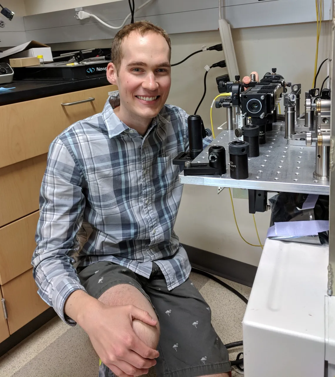
Esophageal cancer, he continues, is particularly hard to detect with current methods. “The big problem is that [cancer in the esophagus] doesn’t look very different [from healthy tissue] until it’s too late,” he says. To address that problem, Sawyer, his mentor Dr. Jennifer Barton, and their colleagues are building a better endoscope, which is essentially a tiny camera on the end of a flexible rod. At the moment, this means experimenting with the ways tissues, both healthy and diseased, react to different types of light.
Sawyer lays out the basics of his research:
What I’m looking at is how the colors interact with the tissue. When you illuminate tissue, it might undergo fluorescence or absorption, which can then give you information about the blood, proteins, and amino acids, and all of these things change in a body fighting a disease. Knowing normal baseline reactions and then measuring changes can give a physician a lot more information than simply looking at the tissue.
These methods, he hopes, will lead to earlier detection and, thus, to lives saved. Additionally, he wants to create new technologies that are more cost-effective and therefore more accessible to patients of all income levels.
Sawyer’s human-centric, community-oriented aspirations are exciting, especially coupled with his obvious passion for improving medicine through improved tools and methods. The future of his work, and all its implications, certainly is bright.

